A Step-by-Step Guide to Nucleic Acid Electrophoresis
Mar 30, 2025
Introduction
Nucleic acid electrophoresis is a fundamental technique in molecular biology used to separate DNA or RNA fragments based on their size. This method relies on an electric field to move negatively charged nucleic acids through a gel matrix, typically agarose or polyacrylamide. The separated fragments can then be visualized and documented using a gel documentation system, such as Yanbiotech’s YB-GD1, for accurate analysis. Below is a detailed step-by-step protocol for performing nucleic acid electrophoresis.
Materials Required
1. Gel preparation:
Agarose powder (for agarose gel electrophoresis)
TAE (Tris-Acetate-EDTA) or TBE (Tris-Borate-EDTA) buffer
Gel casting tray and comb
Ethidium bromide (EB) or safer alternatives like SerRed.
2. Sample preparation:
DNA/RNA samples
Loading dye
DNA ladder (size standard)
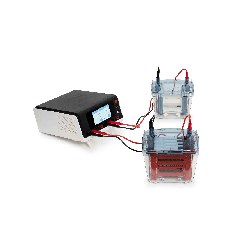
3. Electrophoresis setup:
Electrophoresis chamber
Power supply
Yanbiotech YB-GD1 Gel Documentation System (or UV transilluminator for visualization)
Step-by-Step Procedure
1. Preparing the Agarose Gel
1.1: Prepare the buffer (usually 1X TAE or TBE).
1.2: Weigh the appropriate amount of agarose (e.g., 1% agarose for standard DNA separation).
1.3: Mix agarose with buffer and heat until fully dissolved (microwave for 30-60 sec).
1.4: Cool the solution slightly (~60°C), then add DNA stain (e.g., EB or SerRed).
1.5: Pour the gel into a casting tray with a comb inserted and let it solidify (~20-30 min).
2. Loading the Samples
2.1: Carefully remove the comb and place the gel in the electrophoresis chamber.
2.2: Fill the chamber with enough buffer to cover the gel.
2.3: Mix DNA samples with loading dye (for tracking migration).
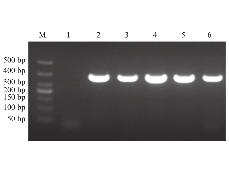
2.4: Load the samples into the wells, including a DNA ladder for size reference.
3. Running the Electrophoresis
3.1: Connect the electrodes (negative to the sample side, positive to the opposite end).
3.2: Apply voltage (typically 80-120 V for agarose gels).
3.3: Run until the dye front migrates ¾ of the gel length (~30-60 min, depending on fragment size).
4. Visualizing and Documenting Results with YB-GD1
4.1: Turn off the power supply and carefully remove the gel.
4.2: Place the gel in the Yanbiotech YB-GD1 Gel Documentation System for high-resolution imaging.
The YB-GD1 offers UV and white LED excitation, allowing visualization of SerRed, and other common nucleic acid stains.
Its high-sensitivity CCD camera ensures clear and precise band detection.
The accompanying software enables automatic band analysis and size estimation by comparing samples to the DNA ladder.
4.3: Save and export the gel image for further documentation and publication.
Troubleshooting Tips
Smearing: May indicate degraded DNA or too much sample loaded.
No bands: Check staining method, electrophoresis conditions, or DNA quantity.
Uneven bands: Ensure gel was poured evenly and buffer concentration was correct.
Poor image quality (if using YB-GD1): Adjust exposure time or focus settings in the software.
Conclusion
Nucleic acid electrophoresis is a simple yet powerful technique for analyzing DNA and RNA. By following these steps carefully and using Yanbiotech’s YB-GD1 Gel Documentation System, researchers can achieve high-quality visualization, accurate sizing, and professional documentation of nucleic acid fragments. This method is widely used in cloning, PCR verification, and genetic fingerprinting, making it essential for molecular biology laboratories.
Why Choose Yanbiotech’s YB-GD1?
✔ High-resolution imaging for precise DNA/RNA band detection
✔ UV and LED options for compatibility with multiple stains
✔ User-friendly software for automatic band analysis
✔ Compact and efficient design, ideal for lab workflows
Would you like any further modifications or additional details on the YB-GD1 system?


 Network Supported Xml / Privacy Policy
 English
 English
 Русский
 Español