
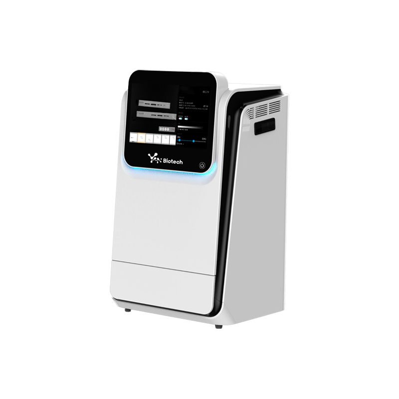
Chemiluminescence Imaging System for Protein Electrophoresis ELC Western Blot Chemi Doc Chemiluminescence Imager
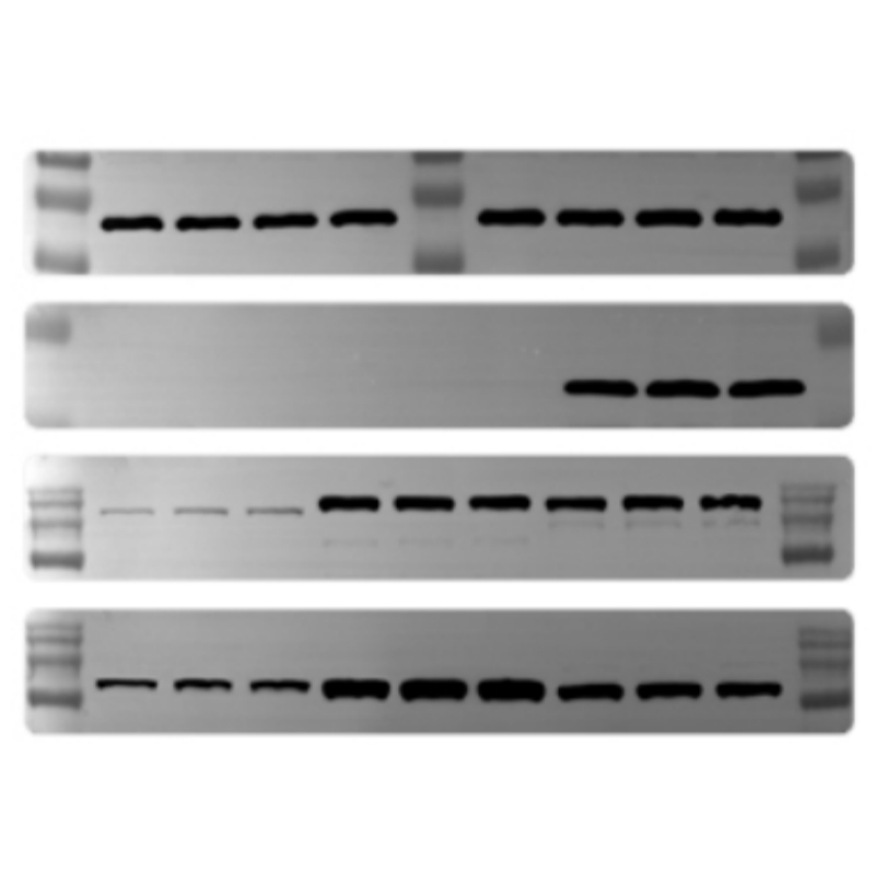
Western blotting, Optional fluorescence application
Cat. No. :
YB-CD2Brand :
YanbiotechProduct Origin :
ChinaSpec. :
Sony ICX695 CCD SensorProduct Introduction
Feature
Application:
Product Specifications
1. CCD Camera
2. Lens
2.1 F0.95 fast lens (fixed Apture)
2.2 Fixed single sample tray, and no focus is necessary
3. Darkroom
3.1 No light leakage darkroom,Size 35x32x50cm
4. Epi-Light Source
4.1 Epi- White LED*2
4.2 Optional EPI-RGB light source for fluorescence application
5. Filter System
5.1 Optional filters for fluorescence application (Alexa 488, Alexa 546, Alexa 647, Cy2, Cy3 and Cy5 as usual specification)
6. Sample tray
6.1 Chemiluminescence
6.2 Optional Fluorescence
7. Optional Removable PC with touch screen
7.1 CPU: Intel I5
7.2 SSD; 128GB
7.3 RAM: 4 GB
7.4 Touch Screen (10.1 Inch)
7.5 Windows 10 Operation System
8. Image Acquisition Software
9. Image Analysis Software
10. Application
10.1 Chemiluminescence (Blotting membrane)
10.2 Optional Fluorescence Applications
Applications


Stay Connected With Us


Copyright @ Wuhan Yanbiotech Co., Ltd. All Rights Reserved.
 Network Supported Xml / Privacy Policy
 English
 English
 Русский
 Español