
GMCSF, CSF2
Cat. No. :
YB3962HBrand :
YanbiotechProduct Origin :
ChinaVolume :
96TSpec. :
Human Elisa KitI. Product Information:
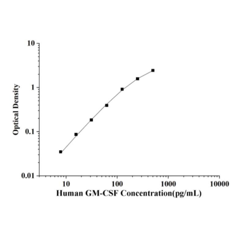
This ELISA kit utilizes a sandwich enzyme-linked immunosorbent assay (ELISA) methodology. The pre-coated microplate wells contain an immobilized capture antibody specific to Human GM-CSF. Following the addition of standards or test samples, the antigen binds to the coated antibody. Subsequently, a biotin-labeled detection antibody specific to Human GM-CSF is introduced, followed by an avidin-horseradish peroxidase (HRP) conjugate. After incubation and washing to remove unbound materials, substrate solution is added to each well. A blue color develops only in wells where the complete immunocomplex (Human GM-CSF, biotinylated detection antibody, and avidin-HRP conjugate) is present. The reaction is terminated by adding stop solution, turning the color yellow. Absorbance is measured at 450 nm ± 2 nm, with optical density (OD) values being directly proportional to the concentration of Human GM-CSF. Sample concentrations are then determined by plotting their OD values against a standard curve.
| Cat Number | YB3962H |
| Alternate Names | GMCSF, CSF2 |
| Detection Method | Sandwich |
| Detection Range | 7.82-500pg/mL |
| Uniprot ID | P04141 |
| Applications | Tumor immunity |
| Species | Human |
| Sensitivity | 4.69 pg/mL |
| Standard | 500pg/mL |
| Sample Type | Serum, Plasma, Tissue homogenate and Other biological samples;Sample Volume=100μL |
| Reaction Time | 3.5H |
II.Sample Testing Protocol:
1. Fresh samples without long time storage is recommended for the test. Otherwise, protein degradation and denaturalization may occur in those samples and finally lead to wrong results.
2. We are only responsible for the kit itself, but not for the samples consumed during the assay. The users should calculate the possible amount of the samples used in the whole test. Please reserve sufficient samples in advance.
3. The detection range of the kit is not equivalent to the concentration range ofthe analyte in the sample. It is recommended to consult reference, conduct preliminary experiments, or seek technical support to estimate the concentration of the analyte in your sample. If the analyte concentration in thesample is too high or too low, appropriate dilution or concentration of thesample should be performed.
4. If the sample type is not included in the manual, a preliminary experiment issuggested to verify the validity.
III. Reagent preparation:
1. Bring all reagents to room temperature(18-25℃) before If the kit will not be used up in one assay, please only take out the necessary strips and reagents for present experiment, and store the remaining strips and reagents at required condition.
2. Wash Buffer: Dilute 30 mL of Concentrated Wash Buffer with 720 mL of deionized or distilled water to prepare 750 mL of Wash Buffer. Note: if crystals have formed in the concentrate, warm it in a 40℃ water bath and mix it gently until the crystals have completely dissolved.
3. Standard working solution:
① Centrifuge: Centrifuge the standard at 10,000×g for 1 min.
② Add 1mL of Reference Standard &Sample Diluent, let it stand for 10 minand invert it gently several times. After it dissolves fully, mix it thoroughly with a pipette. This reconstitution produces a working solution of 2000pg/mL.
③ Serial dilution:Take 7 EP tubes,adding 250μL of reference standard&sample dilution buffer to each tube(The volume can be adjusted based on actual usage,e.g.,500μL/tube). Transfer 250μL of the 2000pg/mL standard working solution into the first tube and mix thoroughly to obtain the 1000pg/mL standard working solution. Continue the dilution step by step until the second-to-last tube. The last tube will serve as the blank, and no solution should be transferred from the second-to-last tube. The standard working solution should be freshly prepared and used immediately.
4. Biotinylated Detection Ab working solution: Calculate the required amount before the experiment(100μL/well). In preparation, slightly more than calculated should be prepared. Centrifuge the Concentrated Biotinylated Detection Ab at 800×g for 1 min, then dilute the 100×Concentrated Biotinylated Detection Ab to 1×working solution with Biotinylated Detection Ab Diluent (Concentrated Biotinylated Detection Ab: Biotinylated DetectionAb Diluent=1:99).
5. HRP Conjugate working solution: Calculate the required amount before theexperiment (100μL/well). In preparation, slightly more than calculated shouldbe prepared. Centrifuge the Concentrated HRP Conjugate at 800×g for 1 min, then dilute the 100×Concentrated HRP Conjugate to 1×working solution with HRP Conjugate Diluent (Concentrated HRP Conjugate:HRP Conjugate Diluent=1:99).
6. Experimental Operation Tips
① Solutions should be added to the bottom of the micro ELISA plate well, avoid touching the inside wall and causing foaming as much as possible.
② Make the tested strips in use immediately after the wash step. Do not allowwells to be dry.
③ After adding the Substrate Reagent. The reaction time can be shortened orextended according to the actual color change, but not more than 30 min.
④ Adding the stop solution should be done in the same order as the substrate solution.
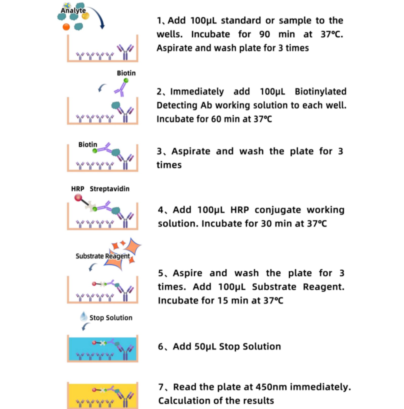
IV. Assay Procedure:
V. Notice:
For Life Science Rearch Only


Stay Connected With Us


Copyright @ Wuhan Yanbiotech Co., Ltd. All Rights Reserved.
 Network Supported Xml / Privacy Policy
 English
 English
 Русский
 Español