
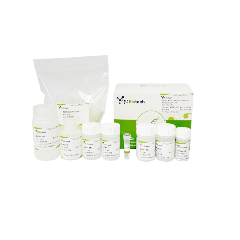
Tissue/Cell/Blood Genomic DNA Extraction Kit Laboratory Research Reagents Biological Chemical Products
Suitable for DNA Extraction from Animal Tissues, Cells and Blood
Cat. No. :
YBD3611-50TBrand :
YanbiotechProduct Origin :
ChinaVolume :
50TSpec. :
Tissue/Cell/Blood GenomicI. Product Introduction
YBD3611-50T, Tissue/Cell/Blood Genomic DNA Extraction Kit utilizes a unique Binding Buffer/Proteinase K system to rapidly lysis cells and inactivate cellular nucleases. Genomic DNA is selectively adsorbed onto the silica membrane of the spin column under high chaotropic salt conditions. A series of quick wash-centrifuge steps using the Inhibitor Removal Buffer and Wash Buffer remove cellular metabolites, proteins, and other impurities. Finally, pure genomic DNA is eluted from the silica membrane with a low-salt Elution Buffer.
II. Product Information:
| Contents | Storage | 50T | 100T | 200T |
| Buffer BL | RT | 5mL | 10mL | 2×10 mL |
| Buffer TL | RT | 11mL | 20mL | 40mL |
| Buffer BB | RT | 10mL | 20mL | 40mL |
| Buffer CB | RT | 15mL | 30mL | 60mL |
| Buffer IR | RT | 25mL | 50mL | 100mL |
| Buffer WB | RT | 15mL | 25mL | 50mL |
| Add the specified amount of ethanol according to the instructions before use | ||||
| Buffer EB | RT | 15mL | 15mL | 2×15mL |
| Proteinase K | -20℃ | 20 mg | 40mg | 80mg |
| Spin EC | RT | 50 pcs | 100 pcs | 2×100 pcs |
| Collection Tube (2mL) | RT | 50 pcs | 100 pcs | 2×100 pcs |
III. Storage Condition:
This kit can be stored for 12 months under dry conditions at room temperature (15°C–25°C),Proteinase K (20mg/mL) need stock in -20℃ individually.
IV . Materials to Be Prepared by the User:
Absolute ethanol, isopropanol, microcentrifuge, water bath.
V. Product Description:
This kit utilizes a unique Binding Buffer/Proteinase K system to rapidly lysis cells and inactivate cellular nucleases. Genomic DNA is selectively adsorbed onto the silica membrane of the spin column under high chaotropic salt conditions. A series of quick wash-centrifuge steps using the Inhibitor Removal Buffer and Wash Buffer remove cellular metabolites, proteins, and other impurities. Finally, pure genomic DNA is eluted from the silica membrane with a low-salt Elution Buffer.
VI . Product Feature:
1. All silica membranes in the spin columns are made from high-quality, specially engineered adsorption material, ensuring minimal variation in binding capacity between columns.
2. The high-quality lysis buffer contains no sodium iodide or perchlorate commonly found in traditional formulations, thus avoiding inhibition of downstream enzymatic reactions such as digestion, ligation, and cloning after recovery.
3. The lysis buffer Buffer is tinted with phenol red, providing a yellow color that facilitates visual monitoring of lysis progress and pH changes, thereby optimizing binding conditions and significantly improving recovery efficiency.
4. The improved lysis buffer Buffer DD formulation greatly enhances buffering capacity and stability, maintaining the pH within the optimal binding range even with highly variable sample types.
5. The procedure is rapid and convenient, eliminating the need for toxic reagents such as phenol and chloroform, as well as ethanol precipitation.
VII. Application:
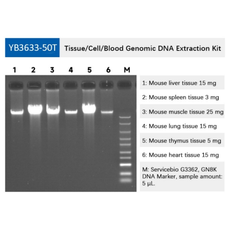
Suitable for rapid extraction of genomic DNA from whole blood and various animal/plant tissues and cells.
VIII. Procedure (Please read the precautions before starting the experiment):
1. Whole Blood
a. Transfer 200 µL of fresh, frozen, or anticoagulated blood to a 1.5 mL microcentrifuge tube.
If the starting volume is less than 200 µL, bring it up to 200 µL with Buffer BB. If the starting volume is between 200-300 µL, proportionally increase reagent volumes in subsequent steps. If the starting volume is between 300 µL-1 mL, perform red blood cell lysis first (see Appendix).
For anticoagulated blood from avian, bird, amphibian, or lower species (which have nucleated red blood cells), use 5-20 µL and bring up to 200 µL with Buffer BB before proceeding.
b. Add 20 µL of Proteinase K (20 mg/mL) solution, mix thoroughly. Then add 200 µL of Binding Buffer CB, vortex immediately and vigorously, and incubate at 70°C for 10 min. The solution should become clear (though may appear dark).
Optional step (usually not required): To remove residual RNA, add 20 µL of RNase A (25 mg/mL) before adding Binding Buffer CB. Vortex, mix, and incubate at room temperature for 5-10 min.
c. After cooling, add 100 µL of isopropanol and vortex immediately and vigorously. Flocculent precipitate may appear.
Immediate and thorough vortexing/mixing in the above steps is critical. Inadequate mixing severely reduces yield. If the sample is viscous, vortex for up to 15 seconds.
d. Transfer the entire mixture (including any precipitate) to a Spin Column AC (placed in a collection tube). Centrifuge at 13,000 rpm for 60 sec. Discard the flow-through.
e. Proceed to step 5 in the General Protocol section below.
2. Tissue Culture Cells
a. Collect approximately 105-106 suspension cells into a 1.5 mL tube. For adherent cells, detach using trypsin before collection.
b. Centrifuge at 13,000 rpm for 10 sec to pellet cells. Discard supernatant, leaving the cell pellet and about 10-20 µL of residual liquid.
c. Resuspend the pellet in 200 µL of 1X PBS. Centrifuge at 13,000 rpm for 10 sec. Completely remove the supernatant. Resuspend the cell pellet in 180 µL of 1X PBS.
d. Add 20 µL of Proteinase K (20 mg/mL), mix thoroughly. Add 200 µL of Binding Buffer CB, vortex immediately and vigorously, and incubate at 70°C for 10 min.
Optional step: To remove residual RNA, add 20 µL of RNase A (25 mg/mL) before adding Binding Buffer CB. Vortex, mix, and incubate at room temperature for 5-10 min.
e. After cooling, add 100 µL of isopropanol and vortex immediately and vigorously. Flocculent precipitate may appear.
f. Transfer the entire mixture to a Spin Column AC. Centrifuge at 13,000 rpm for 60 sec. Discard the flow-through.
g. Proceed to step 5 in the General Protocol section below.
3. Animal/Plant Tissue (e.g., mouse liver/brain or plant leaf)
a. Mince 20-50 mg of fresh or thawed tissue into small pieces with a scalpel (increases yield) or grind to a fine powder in liquid nitrogen. Transfer to a 1.5 mL tube containing 180 µL of Tissue Lysis Buffer TL. Mix by pipetting with a wide-bore tip.
b. Add 20 µL of Proteinase K (20 mg/mL) and vortex immediately and vigorously.
c. Incubate the lysate at 55°C for 1-3 hours or until tissue is completely digested, gently inverting the tube several times during incubation.
Optional step: To remove residual RNA, after step c, add 20 µL of RNase A (25 mg/mL), vortex, mix, and incubate at room temperature for 5-10 min.
d. Add 200 µL of Binding Buffer CB, vortex immediately and vigorously, and incubate at 70°C for 10 min.
e. After cooling, add 100 µL of isopropanol and vortex immediately and vigorously. Flocculent precipitate may appear.
f. Using a 1 mL pipette tip, transfer the mixture to a Spin Column AC. If insoluble tissue clogs the tip, wipe the tip on absorbent paper or discard the tip and clump to avoid clogging the column. Centrifuge at 13,000 rpm for 60 sec. Discard the flow-through.
g. Proceed to step 5 in the General Protocol section below.
4. Animal Tissue (Mouse Tail)
a. Mince a 0.2-0.5 cm mouse tail tip (20-50 mg) *(must be within 0-2 cm from the tip for effective lysis)*, or grind to a fine powder in liquid nitrogen. Transfer to a 1.5 mL tube containing 180 µL of Tissue Lysis Buffer TL. Mix by pipetting with a wide-bore tip.
b. Add 20 µL of Proteinase K (20 mg/mL) and vortex immediately and vigorously.
c. Incubate the lysate at 55°C for 3 hours or until completely digested, gently inverting the tube several times.
Optional step: To remove residual RNA, after step c, add 20 µL of RNase A (25 mg/mL), vortex, mix, and incubate at room temperature for 5-10 min.
d. Homogenize the lysate by passing it 2-3 times through a 1 mL disposable syringe (without needle).
e. Add 200 µL of Binding Buffer CB and 100 µL of isopropanol. Vortex immediately and vigorously.
f. Centrifuge at 13,000 rpm for 5 min. Transfer the supernatant to a Spin Column AC. Centrifuge at 13,000 rpm for 30-60 sec. Discard the flow-through.
g. Proceed to step 5 in the General Protocol section below.
Immediate and thorough vortexing/mixing in the above steps is critical. Inadequate mixing severely reduces yield. If the sample is viscous, vortex for up to 15 seconds.
General Protocol:
5. Add 500 µL of Inhibitor Removal Buffer IR to the column. Centrifuge at 12,000 rpm for 30 sec. Discard flow-through.
6. Add 600 µL of Wash Buffer WB (ensure ethanol has been added!). Centrifuge at 12,000 rpm for 30 sec. Discard flow-through.
7. Repeat step 6 with another 600 µL of Wash Buffer WB.
8. Place the Spin Column AC back into the empty collection tube. Centrifuge at 13,000 rpm for 2 min to dry the membrane completely and remove residual ethanol.
9. Transfer the Spin Column AC to a clean 1.5 mL tube. Apply 100 µL of pre-warmed (65-70°C) Elution Buffer EB directly to the center of the membrane. Let stand at room temperature for 3-5 min. Centrifuge at 12,000 rpm for 1 min. For higher yield, reload the eluate onto the column, let stand for 2 min, and centrifuge again.
Larger elution volumes increase elution efficiency. For higher DNA concentration, reduce the volume, but the minimum should not be less than 50 µL, as too small a volume reduces elution efficiency and yield.
10. DNA can be stored at 2-8°C short-term. For long-term storage, place at -20°C.


Stay Connected With Us


Copyright @ Wuhan Yanbiotech Co., Ltd. All Rights Reserved.
 Network Supported Xml / Privacy Policy
 English
 English
 Русский
 Español